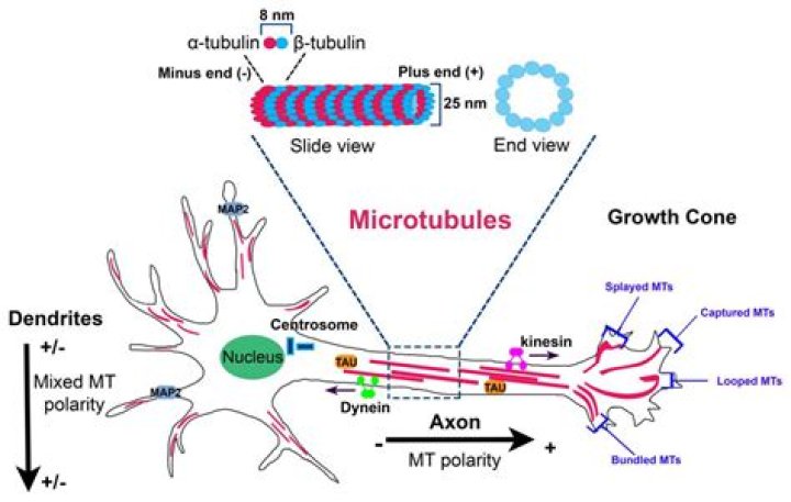
Microtubules (MTs) are long cylindrical structures of the cytoskeleton that control cell division, intracellular transport, and the shape of cells. MTs also form bundles, which are particularly prominent in neurons, where they help define axons and dendrites. Then, what is the main function of the microtubules?
Microtubules have several functions. For example, they provide the rigid, organized components of the cytoskeleton that give shape to many cells, and they are major components of cilia and flagella (cellular locomotory projections). They participate in the formation of the spindle during cell division (mitosis).
Furthermore, what role do microtubules play in neuronal processes? Microtubules play important roles in many cellular functions, including neuronal morphogenesis. During neuronal development, microtubules must form stable bundles, which grow and reorganize to provide the main structural framework for the shafts of axons and dendrites.
Subsequently, one may also ask, what diseases are associated with microtubules?
Reduced microtubule stability has been observed in several neurodegenerative diseases such as Alzheimer's disease (AD), Parkinson's disease (PD), Amyotrophic Lateral Sclerosis (ALS), and tauopathies like Progressive Supranuclear Palsy.
What happens if microtubules collapse?
If a microtubule is severed in its more labile region, the breakage could cause a great deal of disassembly. If the tubulin being yanked is situated at one of the ends of a microtubule, the result would be a shortening of the microtubule from that end; that is, disassembly.
Related Question Answers
What are three functions of microtubules?
Microtubules are part of the cytoskeleton, a structural network within the cell's cytoplasm. The roles of the microtubule cytoskeleton include mechanical support, organization of the cytoplasm, transport, motility and chromosome segregation. What is the structure and function of microtubules?
Microtubules are found in the cytoplasm of all types of eukaryotic cells with rare absence, such as in human erythrocytes. They are tiny, hollow, bead-like tubular structures that help cells maintain their shape. They are microscopic hollow tubes found inside cells that also provide motor functions for the cell. What are the four functions of microtubules?
Functions of Microtubules - Giving shape to cells and cellular membranes.
- Cell movement, which includes a contraction in muscle cells and more.
- Transportation of specific organelles within the cell via microtubule “roadways” or “conveyor belts.”
What is the difference between microtubules and microfilaments?
Microtubules are formed by the polymerization of tubulin proteins. The main difference between microtubules and microfilaments is that microtubules are long, hollow cylinders, made up of tubulin protein units whereas microfilaments are doublestranded helical polymers, made up of actin proteins. What are the different types of microtubules?
There are three main subgroups of microtubules: the polar microtubules (those extending across the cell, as in from centrosome to centrosome), the astral microtubules (those that anchor the spindle poles to the cell membrane), and the kinetochore microtubules (those that extend from the centrosome to the kinetochore What are microtubules made up?
Microtubules are the largest type of filament, with a diameter of about 25 nanometers (nm), and they are composed of a protein called tubulin. Actin filaments are the smallest type, with a diameter of only about 6 nm, and they are made of a protein called actin. What is microtubule dysfunction?
Microtubules are vulnerable in a variety of neurodegenerative diseases. Dysfunctional microtubules are at the center of neurodegeneration and injury. Microtubules undergo complex modifications and protein-protein interactions. Microtubule-mediated mechanisms are ripe with therapeutic targets. What are microtubules quizlet?
maintain axons in nerve cells, mitotic spindles of chromosomes, aid in directional movement of vesicles and organelles. the cytosolic microtubules have jobs such as. cellular movement including cilia, flagella, and basal bodies to which they are attached to. axonemal microtubules have jobs such as. protofilaments. What type of protein is tau?
Tau is a microtubule-associated protein that stabilizes neuronal microtubules under normal physiological conditions. However, in certain pathological situations, tau protein may undergo modifications, mainly through phosphorylation, that can result in the generation of aberrant aggregates that are toxic to neurons. Who discovered centrosome?
The centrosome is the main microtubule organising centre (MTOC) in animal cells and plays an important role in cellular function and regulating cell division. Theodor Boveri first described the centrosome in 1888 and ever since, there has been enormous progress in our understanding of this organelle. What is the function of tubulin?
Tubulin is the protein that polymerizes into long chains or filaments that form microtubules, hollow fibers which serve as a skeletal system for living cells. Microtubules have the ability to shift through various formations which is what enables a cell to undergo mitosis or to regulate intracellular transport. What is the role of Microfilaments?
Microfilaments. Microfilaments, which are the thinnest part of the cytoskeleton, are used to give shape to the cell and support all of its internal parts. What is the main function of ribosome?
Ribosomes have two main functions — decoding the message and the formation of peptide bonds. These two activities reside in two large ribonucleoprotein particles (RNPs) of unequal size, the ribosomal subunits. Each subunit is made of one or more ribosomal RNAs (rRNAs) and many ribosomal proteins (r-proteins). Do human cells have cytoskeleton?
Eukaryotic cells have an internal cytoskeletal scaffolding, giving them their distinctive shapes. The cytoskeleton enables cells to transport vesicles, undergo changes in shape, migrate and contract. Are neurons microtubules?
Neurons are specialized eukaryotic cells that extend long processes to form connections in the nervous system. Like other eukaryotic cells, neurons have a cytoskeleton that consists of three main polymers: microtubules (green), intermediate filaments (purple) and actin filaments (red). What is the function of cytoplasm?
The cytoplasm is the gel-like fluid inside the cell. It is the medium for chemical reaction. It provides a platform upon which other organelles can operate within the cell. All of the functions for cell expansion, growth and replication are carried out in the cytoplasm of a cell. What does kinesin do in the cell?
Kinesins moving along microtubules usually carry cargo such as organelles and vesicles from the center of a cell to its periphery. Dyneins are important in sliding microtubules relative to one other during the beating of cilia and flagella on the surfaces of some eukaryotic cells. What does the cilia look like?
Cilia are slender, microscopic, hair-like structures or organelles that extend from the surface of nearly all mammalian cells. They are primordial. Are microtubules in plant and animal cells?
While both animal and plant cells have microtubule organizing centers (MTOCs), animal cells also have centrioles associated with the MTOC: a complex called the centrosome. Animal cells each have a centrosome and lysosomes, whereas plant cells do not. Where is dynein found?
Dynein is a minus-end-directed microtubule motor protein, which transports a variety of intracellular cargo by hydrolysing ATP to power its movement along microtubule tracks. Axonemal dyneins are found cilia and flagella, whereas cytoplasmic dynein is found in all animal cells. How do microtubules work?
Microtubules are stiff tubes, about 25 nm in diameter. During interphase, they serve as tracks on which organelles and the nucleus are positioned by molecular motor proteins. During mitosis, microtubules form a structure called the mitotic spindle which physically segregates the chromosomes into the two daughter cells. How do microtubules move?
The movements of cilia and flagella result from the sliding of outer microtubule doublets relative to one another, powered by the motor activity of axonemal dynein (Figure 11.53). The dynein bases bind to the A tubules while the dynein head groups bind to the B tubules of adjacent doublets. Where are microtubules absent?
<br> a. Nuclear membrane, chloroplasts, mitochondria, microtubules and pili are absent in prokaryotic cells <br> b. Nuclear membrane, chloroplasts, mitochondria, microtubules and pili are present in eukaryotic cells <br> c. Do bacteria have microtubules?
Microtubules have not previously been found in bacteria, and we lack insight into their evolution from the tubulin/FtsZ ancestor. Using electron cryomicroscopy, here we show that the tubulin homologs BtubA and BtubB form microtubules in bacteria and suggest these be referred to as “bacterial microtubules” (bMTs). Which is a characteristic of microtubules?
Microtubules are long thin structures that consist of the protein tubulin and typically have a diameter of about 25 nm. Characteristics of microtubules that are important for their functions include: Long rigid shape - which enables microtubules to support other structures within the cell. Why do microtubules have a seam?
What is the microtubule “seam”? Microtubules are hollow polymers made of α- and β-tubulin. The αβ-tubulin heterodimer is considered the building block of microtubules because these two subunits are tightly bound to each other and do not dissociate under physiological conditions. Do prokaryotic cells have microtubules?
Many cytoplasmic tubules and fibrous structures within the size range of tubulin tubules and tubulin protofilaments exist in prokaryotes (Table 1). Microtubule-like structures have been found in the nitrogen- fixing bacterium Azotobacter agilis (113). Why are the ends of microtubules called the and ends?
Why are the ends of microfilaments and microtubules called "plus ends" and "minus ends"? -Because the actin and tubulin monomers that make up microfilaments and microtubules have evolved more rapidly than most other proteins. Are microtubules spindle fibers?
Long protein fibers called microtubules extend from the centrioles in all possible directions, forming what is called a spindle. Some of the microtubules attach the poles to the chromosomes by connecting to protein complexes called kinetochores.