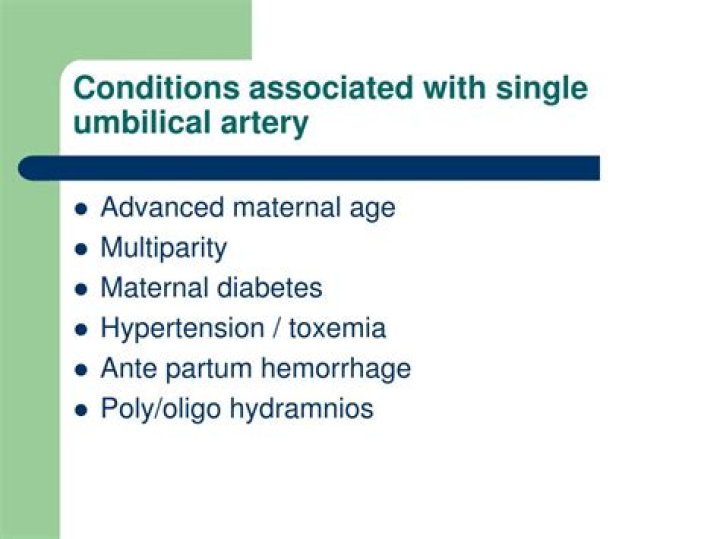
Increased rates of placental, cord, and amniotic fluid abnormalities have been found to be associated with single umbilical artery, including placental structural abnormalities, previa, abruption, abnormal cord length, and hydramnios and oligohydramnios. Accordingly, is single umbilical artery high risk?
Key message. Isolated single umbilical artery is associated with increased risk of adverse perinatal outcome and third stage of labor complications, and there is a risk of recurrence. This population study found that isolated single umbilical artery justifies follow up of fetal wellbeing and labor surveillance.
One may also ask, should I worry about SUA? Pregnancy complications: Another concern with SUA is a possible chance for problems later in pregnancy, like slow fetal growth, preterm delivery, or stillbirth. However, not all studies agree that there is a higher risk for pregnancy complications. Your OB provider will routinely monitor the growth of the baby.
Also question is, should I worry about single umbilical artery?
There are many babies that have a single umbilical artery that have healthy pregnancies and deliveries. However, some babies with a single artery are at increased risk for birth defects.
What happens if baby has single umbilical artery?
Risks Associated with Single Umbilical Artery
We need to maintain a low thresold for diagnosing and managing urinary tract artery infection in there babies. There is an increased risk of fetal growth restriction, prematurity, intra uterine and intra partum death among fetures with single umbilical artery .
Related Question Answers
Is single umbilical artery a birth defect?
A single umbilical artery (SUA) is a malformation of the umbilical cord where only one artery instead of two is present. It may be associated with other birth defects. The pathogenesis of an SUA is thought to be secondary to vessel atrophy of a previously normal cord in the mid trimester. Does Sua mean Down syndrome?
Trisomy 13, trisomy 18, trisomy 21 (Down syndrome), and Turner syndrome are all associated with single umbilical artery, with trisomy 18 being the most common chromosomal abnormality. Are Sua babies born early?
[3] There is also an increased risk of intrauterine growth restriction, prematurity and mortality among neonates with SUA when compared with those with a three-vessel cord. [3,4] This baby was also born premature with a low birth weight. What does Sua mean in pregnancy?
In some cases only a single umbilical artery (SUA) is present. Some report that this umbilical abnormality can be observed as early as 13 weeks. However, it is typically diagnosed in the third trimester. 1,3. There are three theories to explain how a SUA may form during development. Which vein is disappear in umbilical cord?
The expanding amnion surrounds these structures and covers the umbilical cord. Eventually most of the embryonic elements as well as the right umbilical vein disappear, leaving two arteries and one vein (Figure 3.1). Does 2 vessel cord mean Down syndrome?
There used to be a belief that a two-vessel cord signaled an increased risk of chromosomal abnormalities like Down syndrome, but that's actually not true — those would be more likely to be signaled by other symptoms, and your doc would look at other factors to diagnose something like that. What is the function of the umbilical artery?
The umbilical arteries carry deoxygenated blood from fetal circulation to the placenta. The two umbilical arteries converge together about at 5 mm from the insertion of the cord, forming a type of vascular connection called the Hyrtl's anastomosis. What is isolated single umbilical artery?
Single Umbilical Artery (SUA) is characterized by the absence of either the left or right umbilical artery1. This malformation has a reported incidence of up to 1% of pregnancies. The SUA incidence is 3–4 times higher in multiple pregnancies than in singleton pregnancy2. Can Down syndrome be seen at 20 week ultrasound?
A Detailed Anomaly Scan done at 20 weeks can only detect 50% of Down Syndrome cases. First Trimester Screening, using bloods and Nuchal Translucency measurement, done between 10-14 weeks, can detect 94% of cases and Non-invasive Prenatal Testing (NIPT) from 9 weeks can detect 99% of Down Syndrome cases. Can a baby survive placental abruption?
According to the American Pregnancy Association, 15 percent of severe placental abruption cases end in fetal death. Placental abruption is a pregnancy complication that doesn't have a known cause. However, if you're at a higher risk of placental abruption, you can work to reduce your risk. How common are umbilical cord problems?
Nuchal cords are a surprisingly common condition, occurring in as many as 35% of pregnancies. A nuchal cord becomes dangerous when the cord wraps tightly around the baby's neck, often during labor and delivery, and the flow of blood to the baby is interrupted. Can you prevent umbilical cord accident?
There's no way yet to prevent nuchal cords or unwind them from a baby's neck in the womb. But when a baby is born with a nuchal cord, your doctor will know what to do because it happens so frequently. The colored sections of the ultrasound show that the umbilical cord is under the baby's chin. Can you see umbilical cord in ultrasound?
The assessment of the umbilical cord should be an indispensable element of an ultrasound examination in every trimester of pregnancy. The umbilical cord may be visualized for 42 days of gestation as a ropelike, echogenic structure between the fetus and trophoblast [2,3]. Why are there two arteries and one vein in the umbilical cord?
The vein carries oxygen and nutrients from the placenta (which connects to the mother's blood supply) to the baby. The two arteries transport waste from the baby to the placenta (where waste is transferred to the mother's blood and disposed of by her kidneys). What happens to the umbilical artery and vein after birth?
After birth, the proximal portions of the intra-abdominal umbilical arteries become the internal iliac and superior vesical arteries, while the distal portions are obliterated and form the medial umbilical ligaments. The umbilical veins arise from a convergence of venules that drain the extra-embryonic allantois. How common is a 2 vessel umbilical cord?
Two-vessel cord — also known as single umbilical artery (SUA) — occurs in 1 percent of singleton pregnancies and 5 percent of twin and multiple pregnancies. Researchers don't quite know what causes SUA, though they theorize that the second artery disappears at some point during early pregnancy. What is normal umbilical artery PI?
Considering total 2nd and 3rd trimesters the mean PI value of umbilical artery was 1.24 (SD +/- 0.27). While considering the gestational in separate trimesters, study showed that the value of PI in 2nd trimester was 1.33 (SD +/- 0.29) and in 3rd trimester PI was 1.18 (SD +/- 0.25). Is single umbilical artery a soft marker?
[A fetal soft marker in obstetric ultrasonography: single umbilical artery] How can you tell the gender of a baby on an ultrasound?
If the examination of the midline sagittal view of the genital area shows a caudal notch, the fetus is female, and if it shows a cranial notch, then the fetus is male. In the second and third trimesters of pregnancy, ultrasound imaging scans the genital anatomy of the fetus to identify its gender. Is a 3 vessel umbilical cord normal?
The umbilical cord is the connection between your baby and the placenta. A normal umbilical cord has two arteries and one vein. This is known as a three-vessel cord.