Melanocytic hyperplasia (or epidermal melanocytosis) consists in an increased number of melanocytes along the epidermal basal layer. Melanocytes are also more evident for their large size, large nucleus and prominent cytoplasm. Considering this, does melanocytic mean melanoma?
Approximately 33% of melanomas are derived directly from benign, melanocytic nevi. Despite this, the vast majority of melanocytic nevi, which typically form as a result of BRAFV600E-activating mutations, will never progress to melanoma.
Also, what is junctional melanocytic hyperplasia? The histological term, junctional melanocytic hyperplasia (JMH), refers to the proliferation of single melanocytes along the dermal epidermal junction that is the basal layer where normal melanocytes reside. JMH is characteristic of lentigo simplex, junctional nevus, and dysplastic nevus.
In this manner, is melanocytic hyperplasia a cancer?
Atypical melanocytic lesions, which are also called atypical moles or atypical melanocytic hyperlasias, are pigmented moles that contain abnormal cells. These moles are usually benign. However, in some cases, atypical melanocytic lesions can become melanomas.
What is the meaning of melanocytic?
(meh-LAN-oh-site) A cell in the skin and eyes that produces and contains the pigment called melanin. Enlarge.
Related Question Answers
What is the most aggressive form of melanoma?
Nodular melanoma – This is the most aggressive form of cutaneous melanoma. It typically appears as a dark bump – usually black, but lesions may also appear in other colors including colorless skin tones. This type of melanoma may develop where a mole did not previously exist. How long can you have melanoma without knowing?
How long can you have melanoma and not know it? It depends on the type of melanoma. For example, nodular melanoma grows rapidly over a matter of weeks, while a radial melanoma can slowly spread over the span of a decade. Like a cavity, a melanoma may grow for years before producing any significant symptoms. Which stage of melanoma has the best prognosis?
5-year relative survival rates for melanoma skin cancer
| SEER stage | 5-year relative survival rate |
| Localized | 99% |
| Regional | 66% |
| Distant | 27% |
| All SEER stages combined | 93% |
How long does it take for melanoma to spread?
Melanoma can grow very quickly. It can become life-threatening in as little as six weeks and, if untreated, it can spread to other parts of the body. Melanoma can appear on skin not normally exposed to the sun. Where does melanoma usually start?
Melanomas can develop anywhere on the skin, but they are more likely to start on the trunk (chest and back) in men and on the legs in women. The neck and face are other common sites. What do melanomas look like on the skin?
Border that is irregular: The edges are often ragged, notched, or blurred in outline. The pigment may spread into the surrounding skin. Color that is uneven: Shades of black, brown, and tan may be present. Areas of white, gray, red, pink, or blue may also be seen. What is a rare melanocyte?
Rare VariantsThis variant usually presents as indurated plaque or bulky tumor on the head and neck location [60] and is characterized by paucicellular proliferation of atypical dermal spindle melanocytes, dermal collections of lymphocytes with overlying epidermis commonly showing lentigo maligna [61].
What is borderline melanoma?
Background: The borderline melanocytic tumor (BMT) is a morphologically and biologically indeterminate melanocytic proliferation manifesting worrisome architectural features and cytologic atypia exceeding that encountered in melanocytic nevi yet insufficient to warrant designation as melanoma. What causes nodular melanoma?
One main cause is the UV light from the daylight and tanning beds. Their rays can harm the skin's DNA. This damage can happen over many years or even decades. But not all nodular melanomas are caused by UV rays. Is severe dysplastic nevus cancer?
Is it cancer? No. A dysplastic nevus is more likely than a common mole to become cancer, but most do not become cancer. What is Pagetoid spread?
Pagetoid spread is defined as an individual cell proliferation in the upper levels of the epidermis, similar to the pattern of epidermal involvement by Paget's disease of the breast. Is atypical Intraepidermal melanocytic proliferation cancer?
Background: Atypical intraepidermal melanocytic proliferation (AIMP) is a histopathologic diagnosis with uncertain malignant potential and presents a surgical management dilemma for clinicians. What is a regressed melanocytic lesion?
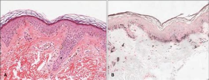
The essential feature of a melanocytic nevus undergoing spontaneous regression is the replacement of tumor cells by a fibrous stroma with varying degrees of inflammation and new blood vessel formation as well as varying numbers of melanophages (Fig. 1). Is severe atypical mole cancer?
About 1 in 10 people develop atypical moles during their lifetime. These moles are not cancerous, and need not be removed if they are not changing. Instead, atypical moles can be a sign of an increased risk for melanoma skin cancer. What is atypical melanocytic nevus?
INTRODUCTION. Atypical nevi, also known as dysplastic nevi, are benign acquired melanocytic neoplasms. Atypical nevi share some of the clinical features of melanoma, such as asymmetry, irregular borders, multiple colors, and diameter >5 mm (picture 1A). They occur sporadically or in a familial setting. What are Monomorphous melanocytes?
Cytologically, the monomorphous melanocytes of melanocytic nevi are in an erratic, unpredictable assortment of small and minimally larger sizes and shapes, e.g., round, oval, spindle-shaped, polygonal, plasmacytoid, ballooned, fusiform, dendritic, pagetoid and multinucleate. Which type of melanoma grows primarily downward into the dermis rather than along the epidermis?
Nodular melanoma is the second most common type of melanoma skin cancer. It makes up about 15% to 20% of all melanoma skin cancers. Nodular melanoma grows down into the skin. It grows and spreads more quickly than other types of melanoma skin cancer. What is Intraepidermal melanocytic proliferation?
Atypical intraepidermal melanocytic proliferation (AIMP) is a descriptive histopathologic diagnosis; the diagnosis at the authors' institution encompasses both likely benign entities (atypical lentigines, lentigines with hyperplasia, and evolving/early junctional dysplastic nevi) and atypical melanocytic lesions that What is compound nevus?
A type of mole formed by groups of nevus cells found in the epidermis and dermis (the two main layers of tissue that make up the skin). What is a compound melanocytic nevus?
Compound melanocytic nevi are simply a combination of junctional and dermal nevi. Nevomelanocytic nests are located at the DE junction in discrete nests, accompanied by an underlying collection of dermal nevomelanocytes (see the image below). What is cytologic atypia?
Cytologic atypia is defined by the presence of nuclei that are large, regularly shaped and hyperchromic, at times showing binucleation and dyskeratosis and presenting with absence of a perinuclear halo. Cytologic atypia can also be found in metaplastic cells. What is dysplastic nevus?
A specific type of nevus (mole) that looks different from a common mole. Dysplastic nevi are mostly flat and often larger than common moles and have borders that are irregular. A dysplastic nevus can contain different colors, which can range from pink to dark brown. What does mild cytologic atypia mean?
Mild cytologic atypia is defined as lesions with ovoid- to ellipsoid-shaped nuclei, that are smaller than basal keratinocytes, with hyperchromatic nuclei, and without a visible or small nucleoli. What are epithelioid melanocytes?
Epithelioid cells are large and round with abundant eosinophilic cytoplasm, prominent vesicular nuclei and large nucleoli. They most commonly arise in superficial spreading and nodular melanomas. Does everyone have the same number of melanocytes?
Special skin cells called melanocytes make melanin. Everyone has the same number of melanocytes, but some people make more melanin than others. The amount of melanin your body makes depends on your genes. What does a melanocytic nevus look like?
They typically appear as small brown, tan, or pink spots. You can be born with moles or develop them later. Moles that you're born with are known as congenital moles. What does a melanocytic nevi look like?
The color ranges from tan to black and can become darker or lighter over time. The surface of a nevus can be flat, rough, raised, thickened, or bumpy; the surface can vary in different regions of the nevus, and it can change over time. The skin of the nevus is often dry and prone to irritation and itching (dermatitis). What does melanoma mean in medical terms?
Listen to pronunciation. (MEH-luh-NOH-muh) A form of cancer that begins in melanocytes (cells that make the pigment melanin). It may begin in a mole (skin melanoma), but can also begin in other pigmented tissues, such as in the eye or in the intestines. How do melanocytes protect the skin?
UVA radiation causes lesions or DNA damage to melanocytes, which are the skin cells that produce the skin pigment known as melanin. Melanin is a protective pigment in skin, blocking UV radiation from damaging DNA and potentially causing skin cancer. Can melanocytes regenerate?
In response to various types of injury, melanocyte stem cells (McSCs) located in the bulge of hair follicles can regenerate mature melanocytes for hair and skin pigmentation. What causes eruptive nevi?
Eruptive nevi are striking — they may appear after a variety of disorders, including Stevens-Johnson/toxic epidermal necrolysis, primary blistering diseases, trauma, immunosuppression (from disease or iatrogenically-induced), PUVA therapy, or idiopathically. Where is melanin produced?
2 Melanin Production. Melanin is produced by melanocytes situated in the basal layer of the epidermis. The melanocortin 1 receptor (MC1R) is regulating the production of both eumelanin and pheomelanin, and the gene encoding MC1R has been sequenced from different ethnic groups (21). What type of cell is a melanocyte?
Melanocytes. Melanocytes are dendritic, pigment-producing cells located in the basal cell layer (Figs 2.4, 2.5). They protect the skin from ultraviolet radiation. Individuals with little or no pigment develop marked sun damage and numerous skin cancers.