Time-lapse fluorescence microscopy is a powerful tool to study gene expression [1]. In this method, protein or mRNA tagged by fluorescence protein (FP) is imaged in real time in individual cells that are actively growing and dividing. Second, this method quantifies fluorescence in single cells. Keeping this in consideration, what is time-lapse microscopy used for?
Time-lapse microscopy can be used to observe any microscopic object over time. However, its main use is within cell biology to observe artificially cultured cells. Depending on the cell culture, different microscopy techniques can be applied to enhance characteristics of the cells as most cells are transparent.
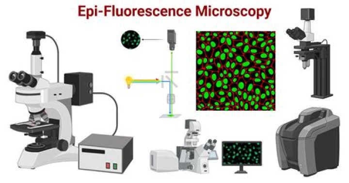
Additionally, what is the principle of fluorescence microscopy? The principle behind fluorescence microscopy is simple. As light leaves the arc lamp it is directed through an exciter filter, which selects the excitation wavelength.
Also question is, what is a time-lapse imaging?
Definition. Time-lapse imaging is a technique whereby serial images are taken at regular time points to capture the dynamics of what is being observed. Recorded images can be played back at different speeds to aid analysis.
How does live cell imaging work?
Live-cell imaging requires a sample in an aqueous environment that is often 50 to 200 micrometers away from the cover glass. Therefore, water immersion lenses can help achieve a higher resolving power due to the fact that both the environment and the cells themselves will be close to the refractive index of water.
Related Question Answers
Which can be used in time lapse microscopy?
Time-Lapse Microscopy - Green Fluorescent Protein.
- Cell Cycle.
- Microscopes.
- Proteins.
- RNA.
- Cell Division.
- Neurons.
- Axons.
What is a confocal microscope used for?
Most confocal microscopes used in industrial applications are reflection-type. They provide a high-resolution image with all areas in focus throughout the field of view, even for a sample having dents and protrusions on the surface. They enable the non-contact non-destructive measurement of three-dimensional shapes. How does phase contrast microscopy work?
Phase contrast microscopy translates small changes in the phase into changes in amplitude (brightness), which are then seen as differences in image contrast. Unstained specimens that do not absorb light are known as phase objects. This allows the specimen to be illuminated by parallel light that has been defocused. What is microscopy in biology?
Microscopy is the technical field of using microscopes to view samples & objects that cannot be seen with the unaided eye (objects that are not within the resolution range of the normal eye). What is the purpose of fluorescence microscopy?
Fluorescent microscopy is often used to image specific features of small specimens such as microbes. It is also used to visually enhance 3-D features at small scales. This can be accomplished by attaching fluorescent tags to anti-bodies that in turn attach to targeted features, or by staining in a less specific manner. What is the use of fluorescence microscopy?
Fluorescence microscopy is a technique used to analyze biological structures in a sample using a white lamp, and either organic or inorganic fluorophores such as dyes to excite a photo-emissive reaction, which is observed using an optical bandpass filter and a dichroic mirror. What is the process of fluorescence?
Fluorescence occurs when an atom or molecules relaxes through vibrational relaxation to its ground state after being electrically excited. The specific frequencies of excitation and emission are dependent on the molecule or atom. What is fluorescence and its application?
Its most common everyday application is in energy-saving fluorescent lamps and LED lamps, where fluorescent coatings are used to convert short-wavelength UV light or blue light into longer-wavelength yellow light, thereby mimicking the warm light of energy-inefficient incandescent lamps. What detector is used in fluorescence microscopy?
Intensified or cooled CCD cameras appear to be the most suitable device for quantitative imaging at low light levels in fluorescence microscopy. Which light is used in fluorescence microscopy?
mercury arc lamp
Does fluorescence microscopy kill cells?
Fluorescence microscopy of fixed cells uses a fixative agent that renders the cells dead, but maintains cellular structure, allowing the use of specific antibodies and dyes to investigate cell morphology and structure. What are the advantages of fluorescence?
Advantages of fluorescence microscopy: Allows labelling of features/molecules of interest and tracking the dynamics of processes involving these features real-time and in vivo. Allows 1–2 magnitude increase in the resolving power of conventional light microscopy, an aspect known as super-resolution microscopy. What is meant by fluorescence?
Fluorescence, emission of electromagnetic radiation, usually visible light, caused by excitation of atoms in a material, which then reemit almost immediately (within about 10−8 seconds). The initial excitation is usually caused by absorption of energy from incident radiation or particles, such as X-rays or electrons. How are fluorescence images recorded?
By scanning the confocal excitation and detection point across the specimen, an image can be sequentially compiled, pixel by pixel, by recording the fluorescence intensity at each position (Fig. The laser excites fluorescence throughout the specimen that passes through the DM and is focused onto the image plane. Which type of microscopy is used for living cells?
phase-contrast microscopy
How do you determine if a cell is living?
A healthy living cell has an intact cell membrane and will act as a barrier to the dye so it cannot enter the cell. A dead cell has a compromised cell membrane, and it will allow the dye into the cell where it will bind to the DNA and become fluorescent. Can fluorescence microscopy be used for live cells?
Fluorescence microscopy of live cells has become an integral part of modern cell biology. Fluorescent protein tags, live cell dyes, and other methods to fluorescently label proteins of interest provide a range of tools to investigate virtually any cellular process under the microscope. Why is cell microscopy important?
A cell is the smallest unit of life. Most cells are so small that they cannot be viewed with the naked eye. Therefore, scientists must use microscopes to study cells. Electron microscopes provide higher magnification, higher resolution, and more detail than light microscopes. Why is live cell imaging important?
Live cell imaging techniques allow the observation of internal structures and cellular processes in real time, and across time. Since live cell imaging is less prone to experimental artifacts, it usually provides more reliable and relevant information than does fixed cell microscopy. What is used to Visualise live cells?
The cells are visualized using a confocal microscope equipped for live imaging or a spinning disc microscope. Fluorescent microscopes can also be used. To reduce the movement of the cells, they can be plated on concanavalin A-coated coverslips. How does ultraviolet light microscopy use fluorescence images?
The ultraviolet light excites fluorescence within molecules in the specimen. This fluorescent light passes through the dichroic mirror and a barrier filter (that eliminates wavelengths other than fluorescent), making it to the eyepiece to form the image. Can electron microscopes view living cells?
Electron microscopes are the most powerful type of microscope, capable of distinguishing even individual atoms. However, these microscopes cannot be used to image living cells because the electrons destroy the samples.